
Skin cancer is the most common form of cancer in humans. The term “cancer” itself refers to an abnormal proliferation of cells resulting in a tumour. These malfunctioning cells can rapidly divide and multiply, and spread to other parts of the body - a dangerous and often fatal process called “metastasis”. Skin cancer occurs when these abnormal cells originate in the layers of the skin, and they are named after the type of skin cell from which they arise. In most cases, as long as the cancerous cells remain in the top layers of the skin, the epidermis, the risk of metastasis and spreading of the cancer to other organs remains low. Once the cancer cells grow deeper into the layers of the skin, the skin cancer becomes more life-threatening. Thus, catching cancer early and preventing the growth of the malignant cells deeper into the skin is essential.
Skin cancers are divided into two main categories: melanoma, and non-melanoma skin cancers. The non-melanoma skin cancers are Basal Cell Carcinomas(BCC) and Squamous Cell Carcinomas(SCC). Together, BCC, SCC and melanoma can take on a variety of shapes and sizes.
Definition
Melanoma is a malignant tumor of melanocytes. Melanocytes are cells that produce melanin, the dark pigments responsible for the color of skin. Melanoma is the most rare, but most dangerous form of skin cancer when detected in a late stage. Fortunately, with early detection the survival rate is 98%.
Signs
Melanoma can appear in moles or spots on the surface of your skin, even in areas not exposed to the sun. In more rare cases it can appear in the eye or bowel. There are four major kinds of melanoma, divided based on clinical and pathological features: superficial spreading melanoma (SSM), nodular melanoma, lentigo malignant melanoma (LMM) and acral lentigious melanoma.
“Melanoma is the most rare, but most dangerous form of skin cancer when detected in a late stage.”
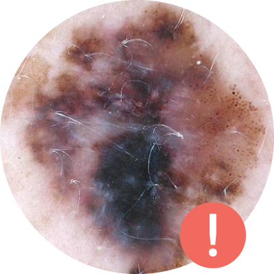
Superficial Spreading Melanoma
This is the most common form of melanoma. Between 70%-80% of melanomas are of this variety, often resembling irregular moles. These melanomas will usually initially grow across the surface of the skin, and are not at risk of spreading and invading other parts of the body until they begin to grow down into deeper layers of the skin. The most common places for these are on the trunk for men and on the legs for women.
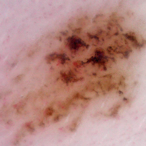
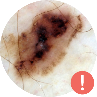
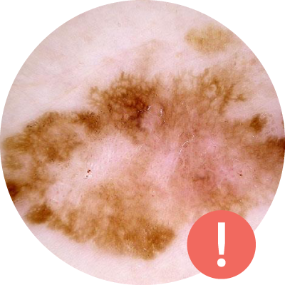
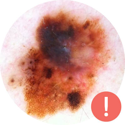
Skin Cancer - Mole Images
Clinical Image
Dermoscopic Image
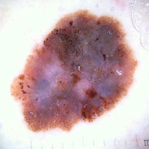
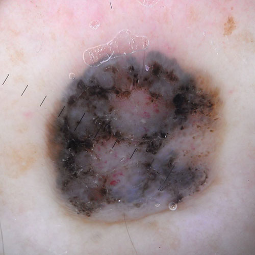
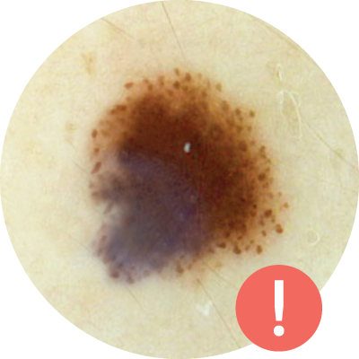
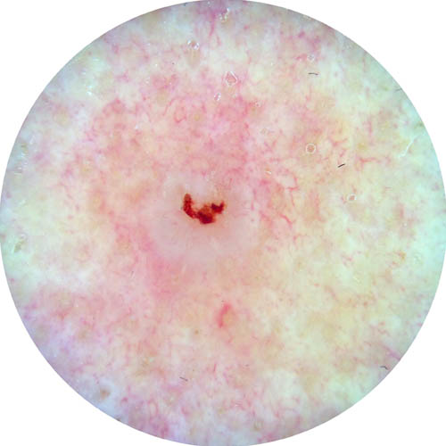
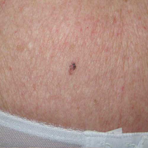
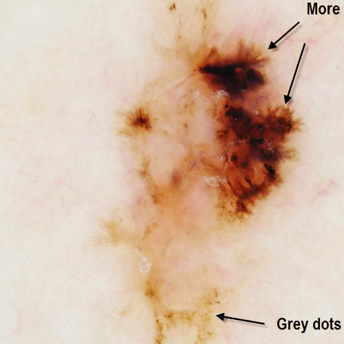
Nodular Melanoma
This form of melanoma grows quickly, and begins growing deep into the layers of the skin sooner than other melanomas. Nodular melanomas often look like black, or very dark brown bumps or raised spots on the skin. These are often found in the middle-aged population on the chest or back.
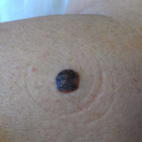
Clinical Image
Dermoscopic Image
Lentigo Malignant Melanoma
This melanoma is similar to superficial spreading melanoma, often remaining on the surface of the skin. It normally develops on sun-exposed areas of the skin, thus it is found on the nose and cheeks, and it is more commonly found in outdoor workers and in older people with sun-damaged skin. Of the 4 types, it is the least likely to become invasive and metastasize.
Clinical Image
Dermoscopic Image
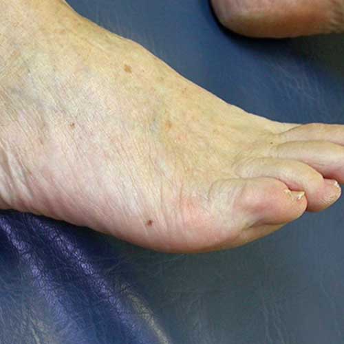
Acral Lentigious Melanoma
This form of melanoma appears under fingernails, on the palms of the hand or soles of the feet. It begins as a slowly-enlarging, discoloured patch of skin that initially grows superficially, but can grow deep into the skin’s layers and become invasive faster than SSM or LMM forms of melanoma. Acral lentigioius melanoma is more common in darker-skinned people, and is often overlooked.
Clinical Image
Dermoscopic Image
What are the ABCDE’s of Melanoma?
Dermatologists use the ABCDE criteria to analyze moles and check for warning signs that may indicate a melanoma.
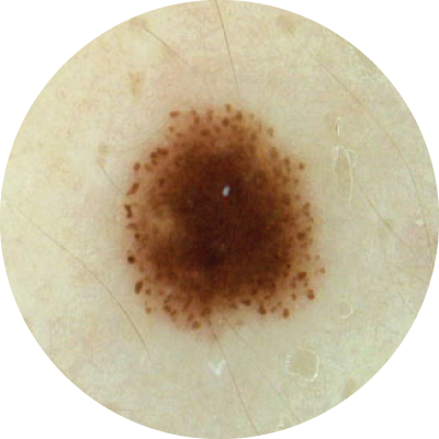
A - asymmetry
Normal moles are symmetrical in shape, meaning if you were to draw a line down the middle, both halves would look the same. Asymmetrical moles are abnormal and should be checked by a doctor.
The image on the left is an example of a symmetrical, benign mole. The image on the right shows what an asymmetrical mole looks like. The two sides of the mole do not match.

B - borders
Blurred, jagged, or irregular borders are a sign that the mole could be cancerous. If the edges of your mole are uneven, it is good to have it checked out by a doctor.
The image on the left shows a mole with defined borders while the mole image on the right has irregular and uneven borders.

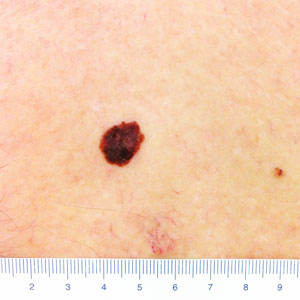
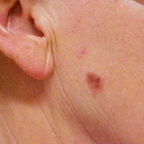
C - colours
Colour can help distinguish a normal mole from an abnormal one. Healthy moles usually have one even colour, while irregular moles can contain multiple shades of colour. It is also important to make note of any blue or white colours that may be present in your moles, as this is a sign that the mole may be cancerous. If your mole has multiple colours or shades, you should speak with your doctor.
The mole in the left image is monotone and is healthy. The image on the right contains multiple shades of red and is cancerous.

D - diameter
In general, if a mole is larger than 6 mm, it is a warning sign, and should be checked by your doctor. As a reference, this is about equivalent to the eraser at the end of a pencil. Even if a mole is smaller than this, it may still be dangerous, so it is important to look for other signs as well.
The image on the right is larger than 6 mm and is cancerous.

E - evolution
Noting any changes with respect to size, shape, colour, or features is an important factor in catching cancer early. If your mole develops symptoms like itching, tenderness or bleeding, or is evolving in any way over time, this is a warning sign to seek medical attention immediately. This is why it is important to not only check your moles regularly, but also to look for any changes that may have occurred since the last self-check you performed.
The image shows the evolution of a mole over time. While not every mole will evolve, or evolve in the same way, even minor changes are important to track and share with your doctor.
Definition
Basal Cell Carcinoma (BCC) is the most common cancer to mankind, with an estimated 2.8 million cases diagnosed each year in the US and 50-60,000 cases per year in Canada . BCC develops from the basal cells in your epidermis. Tanning and UV radiation from the sun contributes to the growth of BCCs, which are commonly found on sun-exposed areas of the body such as the face and neck.
Although it is quite common, BCC is rarely fatal, since it grows slowly, often taking months or years to develop, and is highly unlikely to metastasize (spread to other organs of your body). Nonetheless, a BCC has the tendency to invade into deeper tissue, and thus can be quite disfiguring if it is not properly treated.
Signs
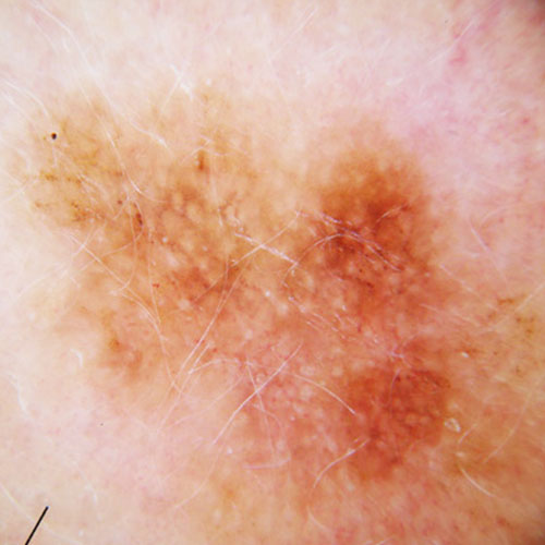
Because of the long growth period, BCCs can vary in size from a few millimeters to several centimeters in diameter. There are several different clinical types of BCCs.
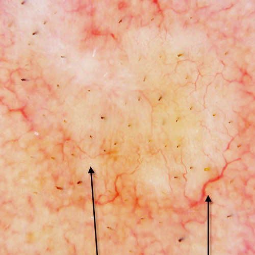
Superficial BCC
- Often multiple pink or red scaly plaques
- Slow-growing
- Commonly found on the upper torso and shoulders
- More common in younger patients
Clinical Image
Dermoscopic Image
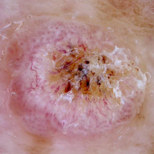
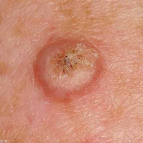
Nodular BCC
- Small, shiny red or pinkish nodule, often with blood vessels (called: telangiectasias) crossing its surface
- May be ulcerated (central open wound)
- Commonly found on the face

Clinical Image
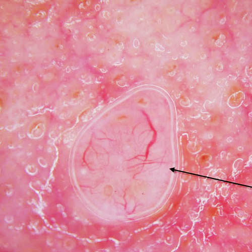
Dermoscopic Image
Morphoeic BCC
- Also known as infiltrating or sclerosing BCC
- Skin-coloured, waxy, scar-like plaques
- More aggressive with poorly-defined edges
- Commonly found on the face
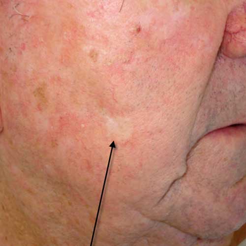
Clinical Image
Dermoscopic Image
Pigmented BCC
- Brown, blue or grayish lesion
- Similar to Nodular or Superficial BCC at the microscopic level
- Can be confused with malignant melanoma
Clinical Image
Dermoscopic Image
Definition
Squamous Cell Carcinoma (SCC) is the second most common type of cancer in humans, with rates as high as 700,000 cases per year in the US and over 20,000 cases each year in Canada. SCC arise from the keratinocyte cells of your epidermis. In many cases, SCC begins with precancerous lesions called actinic keratoses (AKs), which are spots on the skin resulting from years of sun exposure and sun damage. While the majority of AKs either regress or remain AKs, a small percentage of them will turn into SCC. The condition is considered precancerous as long as the damaged cells remain in the epidermis.
If left untreated, the actinic keratosis cells can begin to invade deeper layers of the skin and develop into cancer. Both AK and SCC are most often found on areas of the skin with high exposure to the sun, such as lower lip, exposed scalp, neck, hands, arms, legs and the edges of the ears. Sun exposure is a high risk factor for SCC just like it is for BCC. However, compared to BCC, squamous cell carcinomas are more likely to metastasize and spread to other organs.
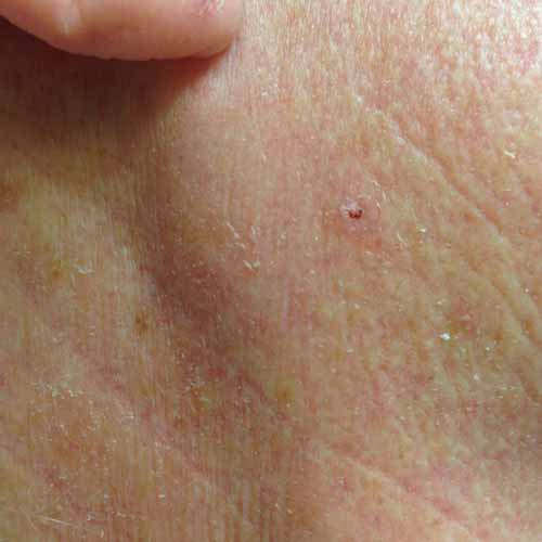
Signs
SCC can present itself in many different ways such as scaly plaques, open sores that may crust or bleed, or even as red patches of skin resembling eczema. Sometimes, SCC can even look like a wart or an elevated growth. Precancerous AK can initially be hard to spot, or nearly invisible, often starting out as a rough patch of skin. AKs are slow-growing, and can eventually appear as a combination of pink, red, dark tan, or skin-coloured spots. They can become inflamed, and in some instances, even bleed. In most cases, AK will regress or remain AK without developing into SCC, however it is important to monitor their development and ensure the condition does not worsen.
Clinical Image